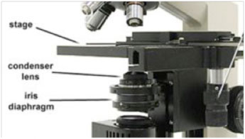
Have you ever wondered how the seemingly simple act of adjusting the light on a microscope can significantly affect the clarity and detail you observe? The answer lies in the often-overlooked yet crucial component known as the diaphragm. This unassuming yet powerful tool plays a pivotal role in shaping the illumination of your specimen, thereby enhancing your microscopic journey.

Image:
The diaphragm, in essence, is a device within a microscope that controls the amount of light passing through the specimen. It is essentially a circular opening that can be adjusted to vary the diameter of the light beam reaching the objective lens. Its importance lies in its ability to optimize image contrast and resolution, allowing you to discern the intricate details of the microscopic world with unprecedented clarity.
Understanding the Mechanics of a Diaphragm
The diaphragm is typically located below the stage, either integrated into the condenser or separately mounted. It consists of a series of interlocking metal plates that can be precisely adjusted to control the size of the aperture. This aperture, in turn, regulates the amount of light reaching the specimen.
There are two main types of diaphragms used in microscopy:
- Iris Diaphragm: This diaphragm uses a lever or knob to open and close the aperture, mimicking the action of an iris in the human eye. It offers smooth and gradual control over the light intensity.
- Disc Diaphragm: This type employs a series of discs with different sized apertures that can be selected by rotating a dial. It provides a set of discrete light settings, offering less fine-tuned control than an iris diaphragm.
The Impact of Diaphragm Adjustment on Image Quality
Adjusting the diaphragm is not a random act. It is a crucial step in obtaining optimal image quality. Here’s how it works:
1. Controlling Light Intensity and Contrast:
As you open the diaphragm, more light reaches the specimen, increasing the brightness of the image. However, too much light can overwhelm the delicate details of the specimen, making it difficult to discern. Conversely, closing the diaphragm reduces the light intensity, improving contrast and making subtle features stand out. This is especially beneficial for observing transparent samples, such as bacteria or protozoa, where the fine details are often masked by excessive light.
2. Optimizing Resolution:
Diaphragm adjustments also affect the resolving power of the microscope – its ability to distinguish between two closely spaced objects. When the diaphragm is fully open, light diffracts excessively, leading to blurring and a loss of detail. By closing the diaphragm slightly, you reduce diffraction and enhance resolution. However, closing the diaphragm too much can create too much contrast, leading to the appearance of artificial edges and a reduction in detail. Finding the sweet spot requires careful adjustment and experience.
Diaphragm Use in Various Microscopy Techniques
The versatility of the diaphragm makes it an indispensable tool in various microscopy techniques:
1. Brightfield Microscopy:
In brightfield microscopy, the diaphragm plays a critical role in controlling the contrast and clarity of the image. By adjusting the aperture, you can balance the light intensity and enhance the visibility of the specimen. This is particularly important when observing stained biological samples.
2. Darkfield Microscopy:
Darkfield microscopy relies on scattered light to illuminate the specimen. In this technique, the diaphragm is closed significantly, blocking most of the direct light. The light then enters the objective lens at an oblique angle, illuminating the specimen from the side. This creates a dark background against which the specimen stands out in bright relief, revealing details often missed in brightfield microscopy. This technique is used extensively for observing transparent specimens, such as bacteria, spirochetes, and fine fibers.
3. Phase Contrast Microscopy:
Phase contrast microscopy, a technique for visualizing transparent specimens, also relies on the diaphragm. In this technique, the diaphragm creates a ring of light that passes through the specimen. The light waves passing through different parts of the specimen interfere, generating contrast based on differences in refractive index. This technique allows scientists to observe the intricate internal structures of living cells without the need for staining.

Image:
Diaphragm Microscopes: Past, Present, and Future
The diaphragm, a fundamental element of microscopy since its inception, has witnessed significant advancements over the years.
1. Evolution of Diaphragm Design:
Early microscopes relied on simple diaphragms with limited adjustability. The advent of the iris diaphragm, offering precise and smooth control, revolutionized microscopy by enabling more detailed and nuanced observations. Modern microscopes often incorporate complex diaphragms that are integrated into the condenser, offering even greater flexibility and precision.
2. The Role of Automation:
In contemporary microscopy, automated systems, such as motorized diaphragms, are becoming increasingly common. These systems offer precise control over light intensity and aperture size, enabling researchers to optimize image quality automatically and with greater accuracy. This automation is especially valuable in high-throughput imaging applications, where consistency and accuracy are paramount.
3. Future Directions:
While the diaphragm remains an essential component of microscopy, ongoing research is exploring new ways to manipulate light and enhance image quality. Future microscopes may incorporate adaptive optics, which dynamically adjusts the light path in real-time, further improving image resolution. Likewise, advances in computational microscopy, such as deconvolution algorithms, are enabling us to extract more information from raw images, surpassing the limitations of conventional optical systems.
The future of microscopy is undoubtedly bright, with the diaphragm playing a crucial role in shaping this exciting landscape.
Diaphragm Microscope
Conclusion
The humble diaphragm, often overlooked, emerges as a powerful tool that shapes the world of microscopy. By understanding its function and mastering its manipulation, you can significantly enhance the quality of your observations. Whether you are an experienced microscopist or a curious beginner, taking the time to appreciate and utilize this indispensable tool will undoubtedly elevate your microscopic journey. So, the next time you are peering into a microscope, don’t forget to give a nod of appreciation to the unassuming diaphragm, the unsung hero of your microscopic exploration.

:max_bytes(150000):strip_icc()/OrangeGloEverydayHardwoodFloorCleaner22oz-5a95a4dd04d1cf0037cbd59c.jpeg?w=740&resize=740,414&ssl=1)


